When the first SARS-CoV-2 genome was published in January 2020, clinical laboratories around the world had a diagnostic test in development within days. That speed was not accidental. It was the product of decades of accumulated expertise in molecular diagnostic techniques, and specifically in the polymerase chain reaction, a method so analytically powerful and technologically adaptable that it has become the backbone of modern infectious disease diagnostics, oncology testing, genetic disease identification, and public health surveillance alike. Molecular diagnostics, the discipline of detecting and characterizing disease through the direct analysis of nucleic acids and proteins, has fundamentally changed what laboratories can know about disease and how quickly they can know it.
The global molecular diagnostics market reflects that reach. Valued at approximately $27 billion in 2024 and projected to grow to $40.4 billion by 2034 at a compound annual growth rate of 4.2 percent, according to Global Market Insights, molecular diagnostic testing now operates across every major disease category, from infectious disease, which held a 71.8 percent share of the market in 2024, to oncology, inherited disorders, and pharmacogenomics. PCR technology alone accounts for 62.16 percent of the molecular diagnostics market by technology share as of 2025, and the PCR-specific segment is projected to grow from $8.8 billion in 2024 to $14.9 billion by 2034. These figures reflect not only the clinical adoption of established PCR platforms but also the accelerating expansion of next-generation methods, from digital PCR and next-generation sequencing to isothermal amplification and CRISPR-based detection systems, that are broadening what molecular laboratories can detect, at what sensitivity, and in what clinical settings.
This article examines what molecular diagnostic techniques are, how PCR works and why it remains central to laboratory practice, and what the expanding toolkit of techniques beyond PCR is contributing to disease diagnosis and patient management.
The Foundation: What Molecular Diagnostics Does
Molecular diagnostic techniques detect disease by identifying specific nucleic acid sequences, DNA or RNA, that are characteristic of a pathogen, a genetic mutation, a chromosomal rearrangement, or an aberrant gene expression pattern. Unlike clinical chemistry, which measures chemical concentrations, or hematology, which examines cell morphology and count, molecular diagnostics works at the level of the genetic code itself. This provides an unparalleled degree of specificity: a well-designed molecular assay can distinguish between two closely related viral strains, detect a single mutated allele against a background of thousands of normal copies, or identify the specific gene fusion driving a tumor’s growth with a precision that no other diagnostic modality can match.
The clinical utility of this precision is extraordinary. Identifying that a respiratory illness is caused by influenza A subtype H3N2 rather than influenza B changes antiviral prescribing. Detecting the EGFR exon 19 deletion in a lung cancer specimen confirms that osimertinib, a targeted EGFR inhibitor, is an appropriate first-line therapy. Quantifying HIV RNA viral load determines whether antiretroviral treatment is suppressing viral replication to undetectable levels or whether resistance mutations may be emerging. None of these clinical questions can be answered by any other laboratory technique with the same combination of specificity, sensitivity, and measurable precision.
PCR: The Engine of Modern Molecular Diagnostics
The polymerase chain reaction, developed by Kary Mullis in the mid-1980s and awarded the Nobel Prize in Chemistry in 1993, is a process for amplifying specific DNA sequences exponentially through repeated cycles of denaturation, primer annealing, and extension by a thermostable DNA polymerase. Starting from as little as a single DNA molecule, a standard PCR reaction can generate billions of copies of a target sequence in a few hours, creating enough material for reliable detection even when the original sample contained only trace quantities of the target.
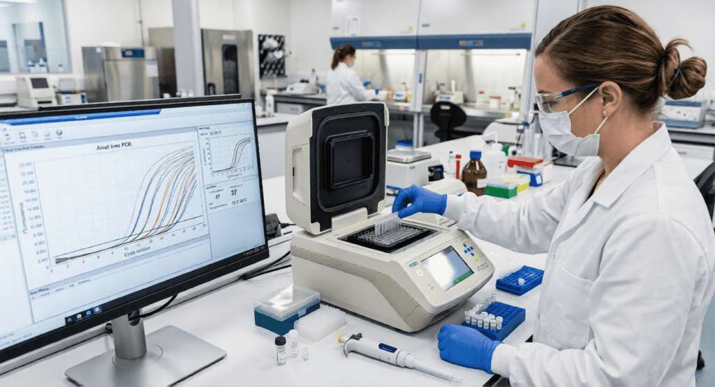
In clinical laboratories, the most widely used PCR format is quantitative real-time PCR (qPCR), which monitors the amplification reaction as it occurs using fluorescent probes or intercalating dyes. The cycle threshold value, the number of amplification cycles required for a sample’s fluorescence signal to cross a defined threshold, is inversely proportional to the amount of target nucleic acid originally present in the sample. This real-time quantification transforms PCR from a qualitative detection method into a quantitative analytical tool capable of measuring viral loads, quantifying gene expression changes, and monitoring treatment response through molecular markers.
For RNA targets, such as RNA viruses including SARS-CoV-2, HIV, and hepatitis C virus, a reverse transcription step precedes the PCR reaction. Reverse transcriptase converts RNA into complementary DNA, which then serves as the template for amplification. Real-time reverse transcription PCR (RT-PCR) is thus the standard method for RNA virus detection in clinical settings.
The diagnostic performance of PCR in infectious disease testing reflects its analytical strengths. During the COVID-19 pandemic, RT-PCR targeting the SARS-CoV-2 ORF1ab, N, and E genes became the global reference standard for diagnosis. Its near-universal adoption was driven by exceptionally high analytical specificity, with well-designed assays showing specificity above 99 percent, meaning false positive rates below one percent even when testing millions of samples. Sensitivity depends heavily on specimen quality and timing relative to the course of infection, since the viral load in nasopharyngeal secretions varies substantially across the illness trajectory. This specimen-dependent sensitivity is a limitation that applies to all nucleic acid amplification tests and explains why negative PCR results in clinically suspicious patients are not definitive exclusions.
In infectious disease testing beyond respiratory viruses, PCR has transformed the diagnosis of conditions where conventional microbiology was too slow, too insensitive, or simply incapable. HIV proviral DNA PCR enables diagnosis of infection in neonates born to HIV-positive mothers before serological responses have developed, a critical capability given that pediatric HIV treatment initiated in the first weeks of life dramatically improves long-term outcomes. Mycobacterium tuberculosis PCR provides results within hours compared to the two to six weeks required for mycobacterial culture, and WHO-endorsed nucleic acid amplification tests such as GeneXpert MTB/RIF simultaneously detect M. tuberculosis and rifampicin resistance mutations, providing both a diagnosis and critical treatment guidance in a single assay within two hours.
The multiplexing capability of modern PCR platforms extends its reach further. In September 2024, Roche launched the cobas Respiratory Panel using its TAGS technology, enabling simultaneous detection of twelve common respiratory viruses, including SARS-CoV-2, influenza A and B, and RSV, on its cobas high-throughput systems. In April 2024, an advanced qPCR test capable of detecting fourteen respiratory viruses simultaneously was launched in Australia by Speed Pty Ltd. These syndromic panels answer a fundamentally different clinical question from single-pathogen assays: rather than confirming a suspected diagnosis, they survey the entire landscape of plausible pathogens from a single specimen, a capability particularly valuable in pediatric respiratory illness where the clinical presentation of viral causes is often indistinguishable.
Digital PCR: Absolute Quantification and Rare Variant Detection

Quantitative PCR provides relative quantification referenced against a standard curve. Digital PCR provides absolute quantification by partitioning the reaction into thousands to millions of individual compartments, each containing zero or one copy of the target sequence, and counting the positive partitions using Poisson statistics. The resulting measurement is a direct molecule count, independent of any calibration curve, and offers substantially greater precision than qPCR at low target concentrations.
Droplet digital PCR (ddPCR), in which the reaction is partitioned into approximately 20,000 water-in-oil droplets, has emerged as the most widely adopted digital PCR format in clinical research and is increasingly entering routine diagnostic practice. The key clinical application where ddPCR’s superiority over qPCR is most consequential is in detecting rare variants at low allele frequencies: mutations present in less than one percent, or even one-tenth of one percent, of the total circulating DNA. These scenarios arise in two critical contexts: liquid biopsy and minimal residual disease monitoring.
In liquid biopsy, ddPCR detects circulating tumor DNA (ctDNA) in blood plasma, where tumor-derived molecules constitute less than 0.1 percent of total cell-free DNA in many early-stage or post-treatment settings. Studies have demonstrated that ddPCR can detect meaningful ctDNA changes in blood several months before imaging scans can detect changes in tumor size, suggesting a future where molecular monitoring outpaces radiological surveillance for early relapse detection. In a Scientific Reports study published in 2024, ddPCR assays for rare BCR-ABL1 fusion transcripts and other leukemia-specific markers showed high sensitivity for minimal residual disease detection in hematological malignancies, with performance assessed through limit of blank, limit of detection, and linear regression analyses confirming the analytical validity of these assays for clinical use.
In minimal residual disease monitoring of hematological cancers, ddPCR has repeatedly demonstrated its ability to detect residual disease below the quantification limit of conventional qPCR. In Philadelphia-positive acute lymphoblastic leukemia, a ddPCR assay for BCR-ABL1 identified quantifiable disease in 53.7 percent of samples that qPCR classified as positive but non-quantifiable, providing clinicians with actionable MRD data that qPCR alone could not supply. In September 2024, QIAGEN released the QIAcuityDx Digital PCR System, a dedicated diagnostic digital PCR platform reflecting the transition of this technology from research settings into certified clinical diagnostic use.
Next-Generation Sequencing: Comprehensive Genomic Profiling in the Clinic
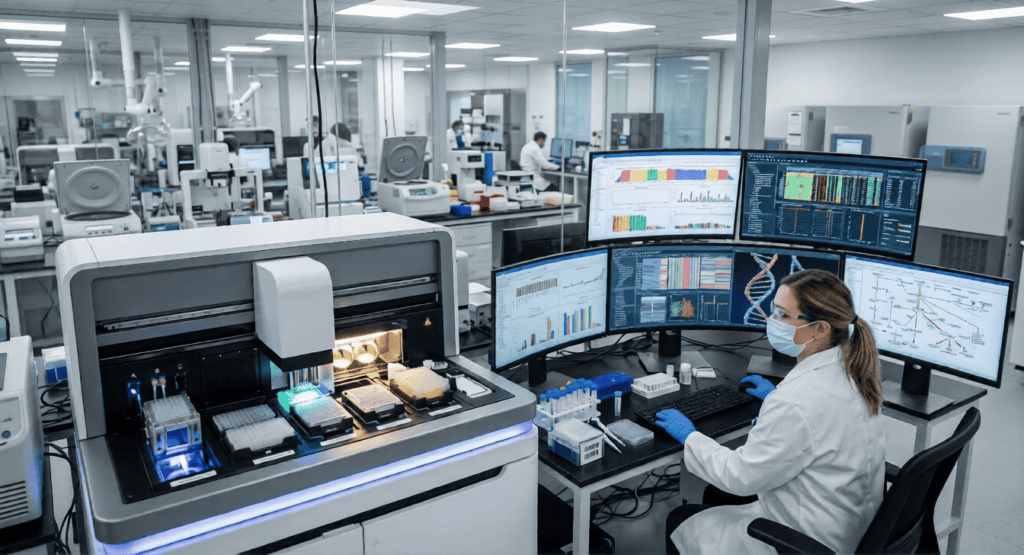
Where PCR targets known sequences with pre-designed primers, next-generation sequencing (NGS) surveys the genome or transcriptome comprehensively, identifying all variants present in a sample without requiring prior knowledge of exactly which mutations to look for. This capability is transformative in oncology, where the genetic landscape of a tumor can include dozens of clinically actionable mutations, copy number variants, gene fusions, and epigenetic alterations, many of which would require separate PCR assays to detect individually.
NGS has changed the way cancer is diagnosed and treated by enabling comprehensive genomic profiling of tumor tissue from a single sequencing run. As of December 2024, over 200 targeted therapies have been approved for cancers defined by specific molecular alterations, from the BCR-ABL inhibitors that transformed chronic myeloid leukemia management to EGFR, ALK, ROS1, BRAF, and KRAS-targeted agents across multiple solid tumor types. The clinical oncology pathway for many of these treatments explicitly requires NGS-based companion diagnostic testing to confirm the presence of the relevant alteration before a targeted therapy can be prescribed.
Beyond treatment selection, NGS contributes to cancer care through detection of hereditary cancer predispositions, where germline sequencing of BRCA1, BRCA2, Lynch syndrome genes, and dozens of others guides surveillance and prophylactic interventions for patients and their families. Tumor mutational burden assessment, which uses NGS to count the total number of somatic mutations per megabase of sequenced DNA, predicts response to immune checkpoint inhibitors across multiple tumor types and is now a recognized biomarker in treatment guidelines from NCCN and ESMO. Spatial transcriptomics and single-cell sequencing, both emerging from NGS infrastructure, are advancing understanding of tumor heterogeneity in ways that promise to further refine which patients respond to which therapies.
The implementation of NGS in clinical laboratories carries requirements that distinguish it from traditional assay validation. Bioinformatics pipelines must be validated alongside the wet laboratory components, and the interpretation of variants of uncertain significance requires specialist knowledge and regularly updated databases. Pre-analytical factors, particularly the degradation of nucleic acids in formalin-fixed paraffin-embedded tissue specimens, introduce errors that can compromise sequencing data quality. Standardization of NGS reporting remains an active area of work for professional organizations and regulatory bodies, as the information density of a comprehensive genomic profile far exceeds that of any single clinical chemistry result.
Isothermal Amplification and CRISPR-Based Detection: Expanding Access Beyond the Central Lab
The analytical power of PCR comes with infrastructure requirements: a thermocycler capable of precise, rapid temperature cycling; a stable power supply; cold chain management for reagents; and trained personnel comfortable with nucleic acid extraction and quality control. These requirements are manageable in well-resourced central laboratories but represent significant barriers in resource-limited settings, field deployments, and point-of-care testing scenarios where results are needed at the bedside or in the community rather than in a laboratory.
Isothermal nucleic acid amplification techniques address these constraints by performing amplification at a constant temperature, typically 37 to 65 degrees Celsius depending on the method, eliminating the need for thermocycling equipment. Loop-mediated isothermal amplification (LAMP) uses six primers targeting eight distinct sequences of the target gene and a strand-displacing DNA polymerase to achieve exponential amplification at a constant temperature with high specificity. Its robustness against common PCR inhibitors found in complex matrices such as blood or feces, combined with the ability to detect results by turbidity or colorimetric change visible to the naked eye, makes it well-suited for settings where laboratory equipment is limited. WHO-endorsed LAMP-based tests for M. tuberculosis and SARS-CoV-2 have been deployed in field settings in low-income countries where conventional PCR infrastructure is unavailable.
CRISPR-based diagnostics, including the SHERLOCK (Specific High Sensitivity Enzymatic Reporter UnLOCKing) and DETECTR platforms, harness the target recognition specificity of CRISPR-Cas systems for nucleic acid detection. When a Cas enzyme loaded with a guide RNA binds its target sequence, it activates a collateral cleavage activity that cuts reporter molecules in the reaction, generating a detectable fluorescent or colorimetric signal. The critical limitation of CRISPR detection without amplification is sensitivity: unamplified clinical samples often contain pathogen nucleic acids at concentrations below the direct detection limit of Cas enzymes. The practical solution, now widely adopted, is to couple CRISPR detection to a preceding LAMP or recombinase polymerase amplification (RPA) step that brings target concentrations into the range where CRISPR can detect them reliably. LAMP-integrated CRISPR platforms combine the speed and temperature simplicity of isothermal amplification with the sequence-level specificity of CRISPR target recognition, addressing the false-positive rate that has been a recognized limitation of LAMP alone. These platforms align with the WHO’s ASSURED criteria for point-of-care diagnostics: Affordable, Sensitive, Specific, User-friendly, Rapid and robust, Equipment-free, and Delivered to those who need it.
Quality Standards and Validation in Molecular Diagnostics

The analytical performance of molecular diagnostic techniques, however sophisticated, depends entirely on the rigor of validation and quality management that surrounds their clinical implementation. Unlike clinical chemistry analytes measured against established reference materials, molecular assays detect sequences whose clinical significance must be established, whose limit of detection must be characterized in the specific specimen matrix used, and whose performance must be monitored through ongoing quality control.
ISO 15189:2022 applies to molecular laboratories as it does to all medical laboratory disciplines, requiring verification of examination procedures, participation in external quality assessment programs appropriate to molecular testing, measurement uncertainty evaluation where applicable, and risk-based thinking about the pre-analytical factors that are particularly consequential for nucleic acid testing: nucleic acid extraction efficiency, inhibitor removal, RNA integrity for transcriptome-based assays, and contamination control in high-sensitivity applications.
The CLSI framework for molecular diagnostics includes specific guidance documents addressing validation requirements for nucleic acid amplification tests, covering analytical sensitivity, analytical specificity, precision, accuracy, reportable range, and reference interval establishment. These validation parameters are not transferable from one laboratory to another even when the same commercial assay is used, because variables including reagent lots, instruments, specimen handling practices, and operator training can all influence assay performance in ways that must be characterized locally.
The Trajectory Ahead
The molecular diagnostics field in 2025 is characterized by three converging trends: the increasing sensitivity of detection methods, enabling earlier disease detection and more granular monitoring; the increasing comprehensiveness of genomic characterization, moving from single-target assays toward whole-genome and multi-omic profiling; and the increasing accessibility of testing platforms, driven by isothermal, CRISPR, and microfluidic technologies that are extending molecular diagnostics toward the point of care and into resource-limited settings where the need for accurate diagnosis is greatest.
The decentralized point-of-care segment of the molecular diagnostics market, while currently smaller than central laboratory testing, is forecast by Mordor Intelligence to grow at the fastest rate of any end-user segment through 2030, with an 8.2 percent CAGR, reflecting the demand for rapid molecular results outside the traditional laboratory setting. As these platforms mature and their quality standards are established, they will bring molecular diagnostic capability to clinical settings and geographic regions where it was previously inaccessible, with direct consequences for the equity of diagnosis across patient populations.
Conclusion
Molecular diagnostic techniques have transformed what clinical laboratories can know about disease and at what speed. PCR remains the dominant technology, its clinical range extending from viral load quantification and pathogen identification to cancer biomarker detection and pharmacogenomic testing. Digital PCR adds the dimension of absolute quantification and ultrasensitive rare variant detection that is redefining cancer monitoring through liquid biopsy. Next-generation sequencing provides comprehensive genomic characterization that is now integral to precision oncology and genetic disease diagnosis. And isothermal amplification coupled with CRISPR detection is extending molecular diagnostics toward settings that PCR infrastructure has never reached.
Across all of these techniques, the same principle applies: the value of molecular diagnostic information depends entirely on the quality with which it is generated, validated, and interpreted. Laboratory professionals who understand not only how to run these assays but what their performance parameters mean and where their limitations lie are the essential human element in a discipline increasingly defined by its technology.
Bio-Reach is a non-profit organization dedicated to advancing Laboratory Medicine through advocacy, education, and global collaboration. To learn more or get involved, visit bio-reach.org.